Aiming to present quantitative indicators that complement morphological observation in rapid on-site evaluation through elemental analysis and nuclear area measurement
Hitachi, Hitachi High-Tech, and the National Cancer Center Hospital have developed a quantitative cellular analysis technology*1 that combines scanning electron microscopy (SEM)*2 and energy-dispersive X-ray spectroscopy (EDX)*3, aimed at application to rapid on-site evaluation (ROSE)*4 in bronchoscopy*5 for peripheral pulmonary lesions suspected of lung cancer.
In conventional ROSE, trained medical professionals use optical microscopes to assess specimen adequacy and to determine benign or malignant status based on morphological observation of cells. The new technology uses the SEM-EDX method*6 to measure phosphorus characteristic X-ray signal counts*7 (hereafter, “P counts”)—which derive from phosphorus (P), a constituent element of DNA—for each cell nucleus. In addition, the area of a cell’s nucleus (hereafter, “nuclear area”) is measured from SEM images. By combining these results, a P count–nuclear area profile can be generated as a quantitative indicator that complements morphological observation. Analysis of the P count–nuclear area profile for bronchial brushing cytology specimens*8 from 49 cases demonstrated that cancer cell groups exhibit distributions distinct from those of normal cell groups. This is expected to support specimen evaluation in ROSE and decision-making regarding the need for additional sampling. Furthermore, by using a compact, easy-to-install tabletop low-vacuum SEM (hereafter, “LVSEM”),*9 the system can be readily introduced even in limited spaces, including examination rooms, and allows direct observation of cytology glass slide specimens without additional preparation or processing.
Going forward, in addition to validating with a larger number of cases, the goal will be to improve measurement and analysis technologies with a view to applying the technology to ROSE in bronchoscopy in order to provide support in clinical practice.

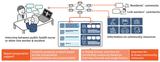
Figure 1: Example of combined use of cytology images (left) and the quantitative indicator P count–nuclear area profile (right) obtained by the SEM-EDX method for application to ROSE (cancer cells are generally characterized by large nuclei and dense nuclear staining)
*1 This technology is in the research stage, and the equipment used in this study has not been approved or certified as a medical device.
*2 Scanning electron microscopy (SEM): An instrument that enables magnified observation of fine structures such as cells.
*3 Energy-dispersive X-ray spectroscopy (EDX): An analytical method used to determine the types and quantities of elements contained in a sample.
*4 Rapid on-site evaluation (ROSE): A method in which cells collected during an examination are evaluated on-site to determine whether there is sufficient specimen for diagnosis and whether additional sampling is needed.
*5 Bronchoscopy: A test performed when opacities are found through CT or other imaging, suggesting lung cancer or infection. A thin endoscope (fiberscope) is inserted through the mouth or nose to directly observe the trachea and bronchi or to collect tissue or cells from lesions (biopsy).
*6 SEM-EDX method: A technique for performing elemental analysis with EDX while simultaneously observing a sample using SEM.
*7 Characteristic X-ray signal counts: In elemental analysis using EDX, the counts of detected X-ray signals characteristic to each element contained in the sample; in this paper, phosphorus characteristic X-ray signal counts are referred to as P counts.
*8 Bronchial brushing cytology specimens: Cell samples collected by brushing during bronchoscopy.
*9 Low-vacuum SEM (LVSEM): A SEM that enables sample observation without conductive treatment such as metal coating. In the newly developed technology, a tabletop low-vacuum SEM is used.
Webpage on the tabletop microscope Miniscope® TM4000PlusIII/TM4000III used in this technology (Hitachi High-Tech)
Webpage on application examples of LVSEM in biopsy tissue (Hitachi High-Tech)
Background and issues
Early detection and improvement in diagnostic accuracy of lung cancer and other respiratory diseases are critical for improving patient survival and clinical outcome. While early detection of lung cancer has progressed with the widespread use of low-dose CT*10 screening in recent years, determining whether small peripheral pulmonary lesions are benign or malignant is difficult based on imaging findings alone, highlighting the need for pathological diagnosis. Bronchoscopy thus requires the collection of specimens suitable for pathological diagnosis, and ROSE is used to assess specimen adequacy in real time during the procedure. Conventional ROSE mainly relies on morphological observation using optical microscopy, in which specialized medical personnel determine specimen adequacy and whether the cells are benign or malignant. However, there has been a demand for the development of methods capable of providing objective quantitative indicators to address cases where determination based solely on morphological observation is difficult.
*10 Low-dose CT: Computed tomography performed using low-dose X-rays. It is a detailed examination method that uses X-rays to obtain cross-sectional images of the inside of the body.
Features of the technology and solutions developed to solve these issues
To offer a quantitative specimen evaluation method for application to ROSE, Hitachi, Hitachi High-Tech, and the National Cancer Center Hospital thus developed a cellular analysis technology that employs the SEM-EDX method. The features of the technology are as follows.
1. P-count measurement technology for objective evaluation of proliferating cells
The SEM-EDX method has made it possible to select regions for each cell nucleus on cytology glass slide specimens and measure P counts. This technology focuses on the possibility that the DNA density within the nucleus increases in proliferating cancer cells, which may be reflected as differences in P counts, which derive from phosphorus (P), a constituent element of DNA. Analysis of cell distribution based on P counts in cytology glass slide specimens showed a statistically significant difference between cancer cell groups and normal cell groups. These results suggest that P counts measured using the SEM-EDX method could be used as an indicator for evaluating proliferating cells.
2. Cellular analysis technology using the P count–nuclear area profile to support specimen adequacy evaluation
Measuring the nuclear area from SEM images and combining it with the P counts obtained above enables cellular analysis based on two quantitative indicators. These indicators may reflect features such as nuclear enlargement and hyperchromasia,*11 which are observed in cancer cells in conventional optical microscopy. By obtaining the P count–nuclear area profile from cytology glass slide specimens and comparing it with a control group of normal cells, this approach holds promise as a quantitative cellular analysis technology that complements morphological observation.
*11 Hyperchromasia: A finding in which the cell nucleus appears densely stained under microscopic observation due to an increase in chromatin content and other factors. In cytology, it is one of the morphological features characteristic of malignant cells.
Confirmed results
For 49 cases of bronchial brushing cytology specimens, P counts and nuclear area were measured for cell groups that physicians had determined to be cancer cells or normal cells based on optical microscopy observation. Depending on the case, cancer cell groups showed significantly higher values in P counts, nuclear area, or both compared to normal cell groups. These results suggest that the cellular analysis technology using the P count–nuclear area profile has the potential to serve as an adjunctive tool for identifying cancer cells. This technology is expected to be used as an indicator that complements morphological observation using optical microscopes in ROSE, thereby contributing to specimen evaluation.
Looking ahead
Going forward, Hitachi and Hitachi High-Tech will continue validation with a larger number of cases. With the eventual application of the technology to ROSE in bronchoscopy in mind, the companies will improve measurement and analysis technologies so that quantitative indicators can be presented in an easy-to-reference format, thereby supporting clinical practice.
Part of these results was published on March 26, 2026, in the international academic journal Scientific Reports.
For more information, use the inquiry form below to contact the Research & Development Group, Hitachi, Ltd. Please make sure to include the title of the article.
https://www8.hitachi.co.jp/inquiry/hitachi-ltd/hqrd/news/en/form.jsp